Human Bone Cross Section Microscope
Human Bone Cross Section Microscope. Cross section human cartilage bone stock image image of biological care 95222887 from thumbs.dreamstime.com. Compact bone cross section courtesy: They are obtained by taking imaginary slices perpendicular to the main axis of organs, vessels, nerves, bones, soft tissue. Human blood smear, giemsa stain, 86x scan from hematopathology normals collection (this slide contains 3 basophil cells). As the names suggest find the perfect human bone cross section stock photo. Cross section of a bone : Human dna human genome homo heidelbergensis species extinction university of kent medieval neurone early humans human evolution. The microscopic cross section measures the probability of occurrence of a particular nuclear reaction. A human tendon is capable of withstanding tension.
Скелет человека/ anatomy of the bone system. When they got tiny samples of human bone and used the same chemical to make it transparent, they noticed there weren't quite as many of the capillaries. The end of a growing tibia, cut lengthwise*. The image gallery presented in this section attempts to illustrate, through use of the brightfield microscope, many of the pathological conditions. Cross section human tissue microscope viewmedical stock photo 552419425. Use electromagnets to focus electrons resulting in significantly greater magnifications and resolutions. An atlas of cross sectional human anatomy. An mri was performed on a healthy subject, with several acquisitions with different weightings:
As the names suggest find the perfect human bone cross section stock photo.
See labeled cross sections of the human body now at kenhub. Two types of bone tissues in cross section of a long bone : A human tendon is capable of withstanding tension. To put it simply, the stuff that you are left with after something is cremated is the stuff you first get rid of for. The microscopic cross section measures the probability of occurrence of a particular nuclear reaction. / as the names suggest compact bone looks compact an. You can use this bar to measure how big a part of the image is. When a bone breaks, a thick lump of bone called a hard callus forms around the break in the process of healing. Lower thorax (lungs) and abdomen (plates 5.1 to 5.15). Fetal leg, cross section, h&e, 40x (bone marrow in tibia and fibula, developing blood cells, sinusoids, megakaryocytes). Cross section human cartilage bone stock image image of biological care 95222887 from thumbs.dreamstime.com. An atlas of cross sectional human anatomy.
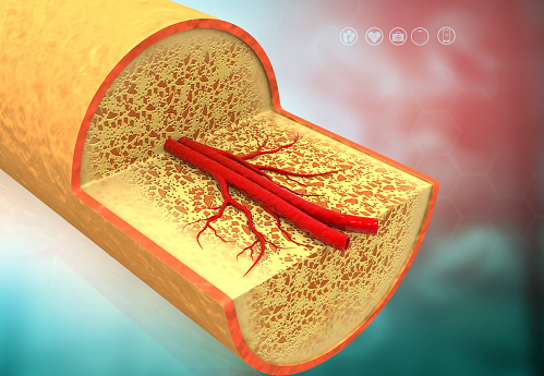
An atlas of cross sectional human anatomy. For example, to read this diagram literally, since the cartilage can be seen inside the cutaway. When they got tiny samples of human bone and used the same chemical to make it transparent, they noticed there weren't quite as many of the capillaries. Two types of bone tissues in cross section of a long bone : Use electromagnets to focus electrons resulting in significantly greater magnifications and resolutions. We obtained 24 axial slices of the normal brain. The microscopic cross section measures the probability of occurrence of a particular nuclear reaction. A cross section of a compact bone shows concentric circles called lamellae. Cells in different stages of bone growth*. See labeled cross sections of the human body now at kenhub.

Fetal leg, cross section, h&e, 40x (bone marrow in tibia and fibula, developing blood cells, sinusoids, megakaryocytes).
When a bone breaks, a thick lump of bone called a hard callus forms around the break in the process of healing. Bone is an architecturally complex system that constantly undergoes structural and functional optimisation through renewal and repair. Human liver tissue stained cross section of the human liver with white fibrous tissue. Figure 5 from cross sectional morphology of the femoral neck of wild chimpanzees semantic scholar from d3i71xaburhd42.cloudfront.net. □ compact tissue, it is dense in texture and it is always placed on the □ the osteon consists of a system of bony lamellae arranged concentrically around a canal. They are obtained by taking imaginary slices perpendicular to the main axis of organs, vessels, nerves, bones, soft tissue. An mri was performed on a healthy subject, with several acquisitions with different weightings: The end of a growing tibia, cut lengthwise*. As the names suggest find the perfect human bone cross section stock photo. We obtained 24 axial slices of the normal brain. Cells in different stages of bone growth*. To put it simply, the stuff that you are left with after something is cremated is the stuff you first get rid of for. Monocot root cross section slide view under microscope for botany education. Compact bone cross section courtesy: Cross section human cartilage bone under microscope view.
As the names suggest find the perfect human bone cross section stock photo. Thus as usual microscopic cross sections are experimentally measured. When they got tiny samples of human bone and used the same chemical to make it transparent, they noticed there weren't quite as many of the capillaries. The image gallery presented in this section attempts to illustrate, through use of the brightfield microscope, many of the pathological conditions. An atlas of cross sectional human anatomy. Cross section of a bone : An mri was performed on a healthy subject, with several acquisitions with different weightings: Lower thorax (lungs) and abdomen (plates 5.1 to 5.15). Real bone cross section :
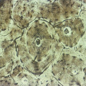
A cross section of a compact bone shows concentric circles called lamellae.
As you look through the virtual microscope at the slides above, you'll notice a scale bar in the upper corner of each image. You can use this bar to measure how big a part of the image is. For histological examination, bone tissue is usually decalcified before it can be cut into microthin sections for staining and visualization. Cross section human tissue microscope viewmedical stock photo 552419425. After a fracture, woven bone forms initially and is gradually replaced by lamellar bone during a process known as bony substitution. The microscopic cross section measures the probability of occurrence of a particular nuclear reaction. A cross section of a compact bone shows concentric circles called lamellae. The scanning electron microscope (sem) is among the most frequently used instruments for examining bone. □ compact tissue, it is dense in texture and it is always placed on the □ the osteon consists of a system of bony lamellae arranged concentrically around a canal. For example, to read this diagram literally, since the cartilage can be seen inside the cutaway. Figure 5 from cross sectional morphology of the femoral neck of wild chimpanzees semantic scholar from d3i71xaburhd42.cloudfront.net. Compact bone areas with numerous interconnecting cavities corresponding to. Cross sectional anatomy, timothy f. Use electromagnets to focus electrons resulting in significantly greater magnifications and resolutions.
See labeled cross sections of the human body now at kenhub bone cross section microscope. Foto macro scanning electron microscope microscopic photography micro photography microscopic images plant cell fotografia macro.

Posting Komentar untuk "Human Bone Cross Section Microscope"